
Abstract. Sleep is not the brain’s off-switch. It is an actively regulated, internally varied state shaped by the interaction of accumulated wakefulness, circadian timing, and distributed neural circuits. This review follows the transition into sleep, the scoring of its stages, the roles attributed to dreaming and memory, lifespan change, major disorders, measurement and treatment, and the debated glymphatic hypothesis. It distinguishes clinical consensus and experimental findings from correlation and speculation, and gives special care to the boundary between animal mechanisms and human evidence.
The central ideas
- Sleep onset reflects an interaction between homeostatic pressure, circadian phase, and neural state-stabilizing systems—not one chemical “sleep switch.”
- NREM and REM labels summarize measurable patterns; stage definitions alone do not prove what a stage is for.
- Dreams are reported from both REM and NREM. Memory benefits are real but selective; a universal emotional “overnight reset” remains unsettled.
- Sleep-duration recommendations are population guidance. Clinical disorders require the right history and, where indicated, validated testing.
- The glymphatic model is an active research hypothesis: animal results are striking, while human studies have not directly established a global overnight waste-clearance rate or clinical benefit.
A guide to the evidence
| Evidence type | What it can support | What it cannot establish by itself |
|---|---|---|
| Consensus scoring or guideline | A standardized clinical rule or recommended care pathway | A complete biological explanation, or the right choice for every individual |
| Controlled experiment | Effects under the tested conditions and outcomes | A universal long-term effect in every population |
| Observational association | Co-occurrence, pattern, or risk signal | That one factor caused the other |
| Animal mechanism study | A tractable biological mechanism in that model | A human rate, diagnosis, or clinical benefit |
| Imaging proxy | A measured signal related to physiology | Direct measurement of an unobserved process unless validated |
Sleep looks like an absence: the lights go out, the body grows still, and conscious contact with the world recedes. Yet the sleeping brain is neither switched off nor uniformly quiet. It cycles through changing electrical patterns, adjusts its responsiveness to the environment, replays and reorganizes some experiences, and coordinates with rhythms that have been evolving since before bedtime. Sleep is better understood as an actively regulated state—one that the brain enters through a negotiated threshold and repeatedly reshapes from within.
That account also needs a caution. Sleep science combines measurements, models, clinical definitions, experiments, and hypotheses, and these do not all carry the same evidential weight. A characteristic EEG pattern is a reliable way to score a stage, but does not by itself explain what that stage is for. An association between sleep and health does not establish that one caused the other. Animal experiments can reveal mechanisms that are difficult to study in people, but cannot simply be translated into a human rate or clinical benefit. Keeping these distinctions visible makes the story more interesting, not less: sleep is a coherent biological architecture whose details are still being mapped.
A night is a sequence, not a single state
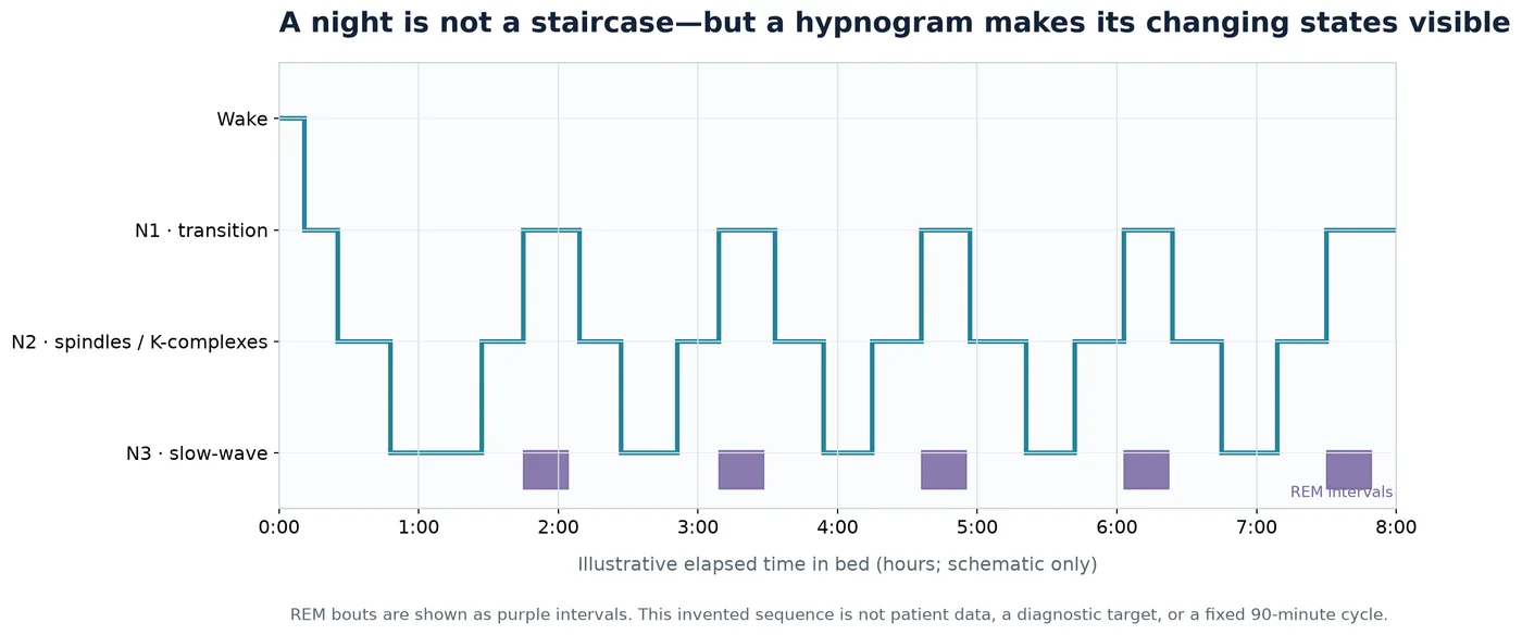
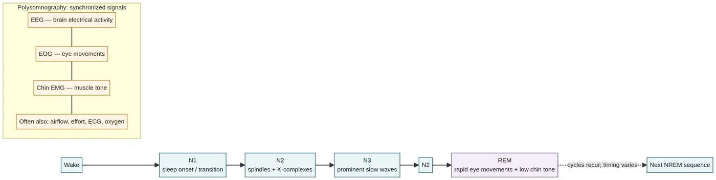
In clinical sleep studies, a night is commonly divided into wake, three stages of non-rapid-eye-movement sleep (N1, N2, and N3), and rapid-eye-movement sleep (REM, also called stage R). These labels are operational: trained scorers use electroencephalography (EEG), eye movements, and muscle activity to classify brief portions of a recording. The American Academy of Sleep Medicine’s current Scoring Manual, Version 3, is the professional reference; the detailed criteria described here are drawn from an accessible earlier version, and the current manual is evolving1,2.
The familiar unit is a 30-second epoch. If more than one stage appears, the scorer assigns the stage occupying the largest portion, subject to rules for particular events and transitions. In wakefulness with eyes closed, an occipital alpha rhythm around 8–13 hertz can be prominent. As sleep begins, alpha activity attenuates or gives way to lower-amplitude mixed-frequency activity; theta-range activity around 4–7 hertz is characteristic of N1. N2 is recognized by features including sleep spindles—brief bursts typically in the 11–16 hertz range lasting at least half a second—or K-complexes, large waveforms with a similar minimum duration. N3 is the consolidated slow-wave stage. REM combines low-amplitude mixed-frequency EEG, rapid eye movements, and markedly reduced chin-muscle tone2,3.
These are not separate rooms with doors that click shut. Brain activity evolves continuously, and a 30-second label compresses fluctuations, brief events, and boundary states into one category. A hypnogram—the chart that plots those categories over time—is useful precisely because it simplifies. It is not a complete portrait of the night. The scorer’s montage, rules, and recording context influence what appears, and laboratory adaptation can alter the pattern. In a two-night study of healthy young adults, stage continuity and cycle-level measures differed on the first, adaptation night4. That result cautions against treating one laboratory hypnogram as an immutable signature of a person.
Over a typical night, NREM and REM recur in several cycles. The often-quoted cycle of roughly 90 minutes and three to five cycles per night are descriptive averages, not a timer built into every brain. N3 tends to be more prominent earlier, while REM periods often lengthen toward morning, but the sequence varies among people and nights3,4. A clinical polysomnogram (PSG) uses EEG, electrooculography (EOG) and chin electromyography (EMG) to stage sleep; it can also record heart activity, airflow, breathing effort, and oxygen saturation. These additional signals answer different questions from staging, such as whether breathing events or oxygen changes accompany the night3.
Sleep stages are a map of patterns, not a ready-made account of their purpose. Slow waves, spindles, eye movements, and muscle atonia are measurable features. Claims about tissue repair, learning, or emotional processing require evidence beyond the definition of the stage itself. That distinction will matter throughout this article: physiology tells us what is happening; experiments and carefully interpreted observations help establish what it may do.
The threshold: homeostatic pressure meets the clock
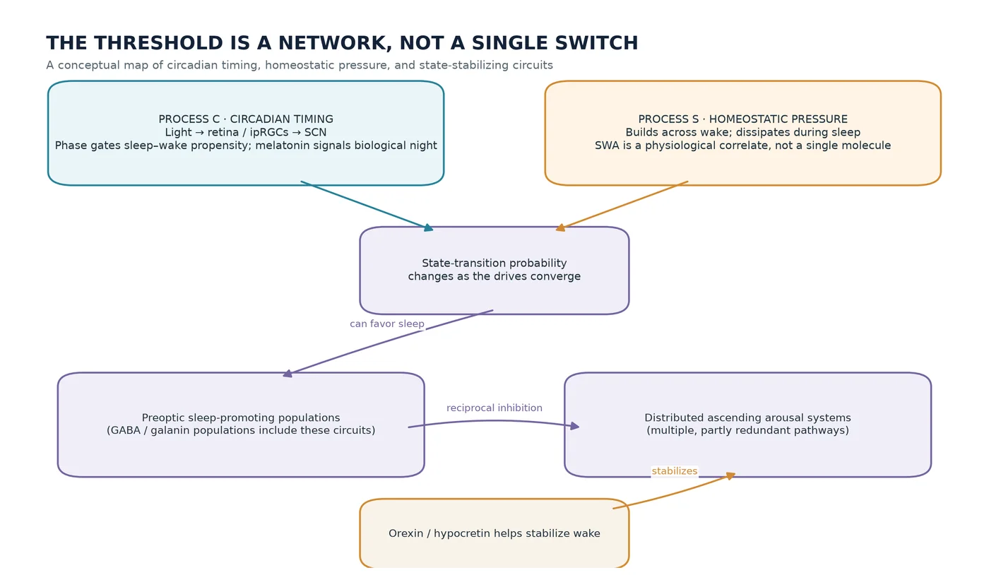
Two broad influences help explain why sleep becomes likely at a particular time. The first is the homeostatic response to time awake. In the classic two-process model, this is Process S: a drive that builds during wake and dissipates during sleep. The second is Process C, the circadian timing signal that changes sleep propensity across the approximately 24-hour day. The model explains much of the broad pattern of sleepiness and alertness, and forced-desynchrony experiments—where schedules are arranged so behavioral timing and the internal clock drift apart—support the idea that homeostatic and circadian effects can be distinguished and interact5.
The model is powerful because it captures a real opposition. After a long waking day, homeostatic pressure tends to rise. Yet the circadian system can provide a late-day or evening signal that temporarily supports wakefulness, helping explain why a person may feel a “second wind” despite having been awake for many hours. At other circadian phases, the same accumulated wakefulness may feel more difficult to sustain. When sleep occurs, homeostatic pressure falls, with the steepest decline in slow-wave activity (SWA), the EEG power associated with large, slow oscillations, often appearing early in the night5.
Process S and Process C are model constructs, not two isolated substances or anatomical modules. SWA is a useful physiological index of homeostatic sleep intensity, not a direct reading of a single molecular reservoir. The model’s equations describe observed time courses; they do not identify one molecule that stores the need for sleep. Cellular and molecular processes interact, and their boundaries do not necessarily match the neat separation of the model5.
Adenosine is one candidate contributor, and caffeine offers an everyday clue: caffeine blocks adenosine receptors, and its alerting effect is consistent with a role for adenosine signaling in sleep-wake regulation. Evidence from pharmacology and animal and human studies supports adenosine’s involvement in adaptive responses to prolonged wakefulness and recovery sleep. But that is narrower than saying adenosine is the sleep-pressure molecule. Measurements show extracellular adenosine can change on a minutes-long timescale, while the buildup and dissipation of SWA unfold over much longer periods. Available profiles therefore do not establish adenosine alone as the generator of ordinary daily sleep pressure6. The likely picture is a network of signals and adaptations, with adenosine participating but not necessarily governing the entire process.
The transition itself can be surprisingly abrupt. Sleep-promoting populations in the ventrolateral preoptic area and median preoptic region, including neurons that use GABA and galanin, inhibit parts of the distributed arousal system. Wake-promoting systems include monoaminergic, cholinergic, and other pathways ascending through the brain. In the “flip-flop” model, reciprocal inhibition between sleep-and wake-promoting populations helps turn gradual changes in sleep pressure and circadian drive into relatively rapid, stable transitions7.
The metaphor is useful, but it should not be mistaken for a literal two-node switch. Neural firing patterns, lesions, and EEG state transitions support important parts of this arrangement; nevertheless, arousal is distributed, pathways overlap, and redundancy is substantial. Lesions of a sleep-promoting region do not abolish all sleep, and some arousal-system lesions have more modest effects than a simple switch diagram might suggest. The model is a compact account of a complex control system, not its final wiring diagram7.
Orexin, also called hypocretin, helps stabilize wakefulness. In animal experiments, activating orexin neurons promotes waking, while inhibiting them promotes sleep. In humans, narcolepsy type 1 is strongly associated with loss of orexin-producing neurons or deficient orexin signaling, convergent evidence that the system normally helps keep wakefulness stable and limits inappropriate intrusion of REM-like features8. Still, the precise contributions of orexin peptides versus the neurons’ other transmitters, the relevant receptor sites, and the dynamics of natural release remain under investigation. The threshold is therefore not a single lever: it is an emergent transition in a distributed system, shaped by accumulated wake, clock time, and interacting neural circuits.
The clock that sets biological night
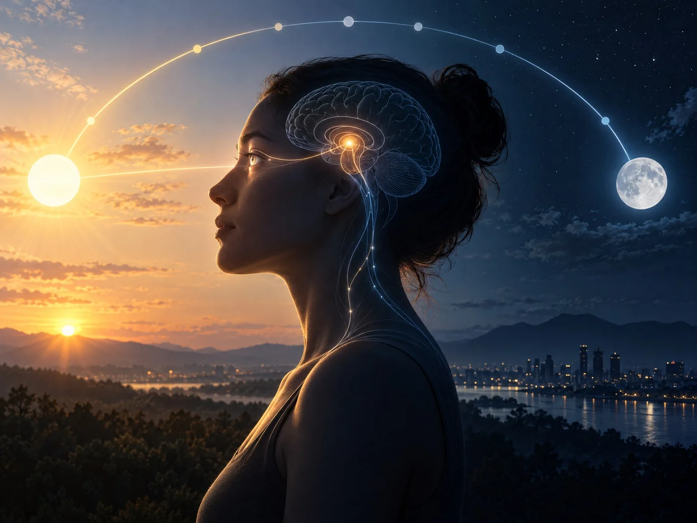
The circadian system keeps time even when schedules and clocks are removed. Its central coordinator in mammals is the suprachiasmatic nucleus (SCN), a small region of the hypothalamus. SCN activity helps organize daily rhythms in sleep propensity, core body temperature, melatonin, and cortisol. In humans, the intrinsic circadian period averages slightly longer than 24 hours, so the clock must be reset by external time cues. The most important cue is light9,10.
Light reaches the SCN through the retina. Melanopsin-containing intrinsically photosensitive retinal ganglion cells (ipRGCs) project directly to it, while rods and cones also contribute to non-image-forming responses. This is why the common shorthand “blue light keeps you awake” is incomplete. Circadian responses depend on wavelength, brightness at the eye, duration, timing within the circadian cycle, prior light exposure, and individual factors. A controlled human study using long monochromatic exposures found that spectral sensitivity changed with exposure duration and that cone input contributed more strongly early in the exposure than later11. The biology is not reducible to a single color or a universal brightness threshold.
Timing determines the direction of a light-induced phase shift. Light in the morning or late biological night tends to advance the circadian clock, while light in the evening or early biological night tends to delay it9. An acute suppression of melatonin and a shift in circadian phase are related but distinct responses. One cannot infer the size or direction of a phase reset simply from how much melatonin was suppressed at a moment9,11.
Melatonin is best thought of as a signal of biological night, regulated by the SCN, rather than a simple sleep switch. The time at which melatonin begins to rise under dim conditions—dim-light melatonin onset, or DLMO—is a useful marker of circadian phase. Core temperature and cortisol also follow daily rhythms, but behavior, meals, sleep, and other factors can mask or alter their observed timing10. A person can be sleepy for homeostatic reasons at a clock time that is not their biological night, or alert at a time when a different schedule would have invited sleep.
The conflict between social schedules and internal timing is sometimes called social jet lag, usually measured as the difference between weekday and weekend sleep midpoints. It correlates with chronotype and has been associated in observational studies with mood, metabolic, and performance measures. In one small study of 24 people, social jet lag was associated with shifts in the timing of selected clock-gene expression peaks12. Neither the association nor the gene-expression finding proves that social jet lag causes disease. Work hours, sleep duration, chronotype, socioeconomic conditions, and other behaviors may contribute.
Shift work makes the mismatch concrete: work can demand alertness during biological night and sleep during the circadian day. This can combine circadian misalignment with insufficient or fragmented sleep and can impair performance. Yet adaptation varies. Some workers shift their circadian timing more than others; schedules, light exposure, days off, and individual biology all matter, and phase alignment alone does not explain every outcome10,13. It is more accurate to describe shift work as a recurring challenge to sleep and circadian organization than as a condition to which every body adapts in the same way.
What the sleeping brain may be doing
Dreams make sleep feel like an interior world, but a dream is not synonymous with REM. Reports of experience can follow awakenings from both REM and NREM sleep. REM reports are more often vivid, perceptually rich, and emotionally intense, but NREM is not a blank interval14,15. Dream research has a built-in measurement problem: experience is usually inferred from a report after awakening. A person’s recall and ability to describe a dream are not identical to the experience itself.
A high-density EEG study repeatedly awakened 14 adults from NREM sleep. Dream reports were preceded by fewer, smaller, shallower slow waves and faster spindles, especially over central and posterior regions14. This links the state of the brain to whether an experience is reported. It does not show that the EEG pattern caused a dream, nor that dreaming performed a particular function. The difference between correlates of reportability and a demonstrated function is essential here.
Sleep’s role in memory is more securely grounded, though still diverse. Across multiple forms of learning, evidence supports reactivation and stabilization of some memories during sleep. The process is not one universal operation, and effects vary with the information learned, task, timing, and sleep physiology16. Staging alone cannot say what a specific memory did during a particular night, and not every memory benefits in the same way.
Emotional memory is a useful example of the limits of broad claims. REM sleep has been proposed as a privileged setting for processing emotional material, in part because REM has characteristic neural and limbic activity. Yet a meta-analysis of 22 sleep-versus-wake comparisons found no overall selective advantage of sleep for emotional over neutral visual recognition memory. A separate analysis of eight stage-specific studies was consistent with REM favoring emotional relative to neutral memory compared with slow-wave sleep, but that smaller evidence base and the specific visual-recognition tasks counsel restraint17. A proposed REM-centered model remains a framework, not a settled rule for all emotional learning15,18.
The stronger-sounding claims—that dreams defuse negative affect, extinguish fear, rehearse coping, or simulate social threats—remain plausible hypotheses with limited direct functional tests15. The narrative content of a dream may be fascinating, but it does not establish that the story itself is doing therapeutic or adaptive work. Sleep may shape memory and emotion through neural processes that do not depend on a dream being remembered, or even experienced as a coherent narrative. That remains an open question.
The changing architecture across a lifetime
Sleep does not have one stable form from infancy onward. Early in life, sleep is distributed across the day and night; over development it tends to consolidate into longer nighttime periods, while naps decline as circadian regulation matures. Nap cessation often occurs somewhere around ages three to five, with substantial variation across children and contexts19. These are population patterns rather than a timetable every child follows.
Consensus recommendations illustrate how strongly sleep duration varies with age. The American Academy of Sleep Medicine recommends 12–16 hours per 24 hours for infants aged 4–12 months, including naps; 11–14 hours for ages 1–2; 10–13 hours for ages 3–5; 9–12 hours for ages 6–12; and 8–10 hours for ages 13–1820. The panel did not set a recommendation for infants younger than four months because normal variation is wide and evidence linking duration to outcomes was insufficient. For adults aged 18–60, a joint AASM/Sleep Research Society consensus recommends regularly obtaining at least seven hours per night21. Those ranges are population guidance, not individualized prescriptions, and sleep quality, timing, regularity, and disorders matter alongside duration. The adult consensus applies specifically to ages 18– 60; older-adult bands published by CDC draw on separate recommendations22.
Architecture changes too. Reviews associate aging with a lower proportion of slow-wave sleep, more N1/N2, and greater wake after sleep onset and fragmentation. Some measures appear to level off in older age groups in reviewed datasets, and results vary by health, methods, and cohort23. This does not mean older people simply need less sleep, or that a particular hypnogram is an inevitable consequence of age. Lifespan research still lacks enough long-term, within-person measurement to clarify how changing sleep and health influence one another over time19.
Habitually insufficient sleep is associated with a range of adverse outcomes, including metabolic and cardiovascular conditions, mood problems, impaired immune function, pain, reduced performance, and accidents21,22. Associations do not mean short sleep alone caused each condition: illness may itself disturb sleep, and lifestyle, work, and socioeconomic factors can confound observed relationships. A critical review of prospective studies cautioned that mortality findings based on self-reported duration are inconsistent and vulnerable to measurement error, response bias, confounding, and reverse causation24.
Experimental sleep restriction offers more direct evidence for some outcomes. In a randomized crossover study, 65 adults experienced six weeks of habitual adequate sleep and six weeks with sleep curtailed by 1.5 hours; performance on working-memory and inhibitory-control/attention tasks was worse under restriction, within the study’s design and sample25. Broader reviews find particularly robust effects on attention and vigilance, with lapses, slowed responses, and unstable alertness; effects on other cognitive domains vary by task and person26. These experiments establish causal effects under defined conditions, not a precise forecast for every individual or every chronic disease.
When sleep’s architecture is disrupted
Sleep disorders are not simply different degrees of “bad sleep.” They are classified by patterns, timing, symptoms, and, where necessary, objective physiology. Insomnia disorder involves persistent difficulty initiating or maintaining sleep, or early awakening, despite adequate opportunity, together with daytime consequences. Modern chronic criteria generally require symptoms at least three nights a week for at least three months; this differs from the older one-month definition in a 2008 guideline27. A short night alone does not establish insomnia, and dissatisfaction without impairment is not the whole syndrome.
Obstructive sleep apnea (OSA) involves recurrent obstruction of the upper airway during sleep. Adult diagnostic criteria cited in AASM guidance include an obstructive respiratory disturbance index (RDI) of at least five events per hour with typical symptoms, or at least 15 per hour regardless of symptoms28. RDI includes respiratory effort-related arousals as well as apneas and hypopneas. The apnea-hypopnea index (AHI) can differ according to how hypopneas are scored—for example, whether a 3% oxygen drop or an arousal counts versus a 4% desaturation requirement. These thresholds are adult classification criteria, not a complete measure of severity or risk, and pediatric criteria differ28.
Circadian rhythm sleep-wake disorders concern persistent misalignment between a person’s endogenous timing and the timing required or desired, producing sleep difficulty or excessive sleepiness and impaired functioning. Sleep may be more normal when the person can follow their biologically preferred schedule. Sleep diaries and actigraphy across time can help characterize regularity and phase10,29. Restless legs syndrome (RLS) is different again: it is a waking sensorimotor pattern involving an urge to move, often with unpleasant sensations, that begins or worsens at rest, improves at least partly with movement, and is worse in the evening or night. Diagnostic criteria also require that symptoms not be better explained by a mimic30.
Narcolepsy is marked by persistent excessive daytime sleepiness. Cataplexy—a brief emotion-triggered loss of muscle tone with consciousness preserved—is characteristic of type 1. Consensus-era objective pathways include a mean sleep latency of eight minutes or less with at least two sleep-onset REM periods on the multiple sleep latency test, or low cerebrospinal-fluid hypocretin-1, interpreted in clinical context31. Sleep paralysis and hallucinations around sleep transitions can occur, but alone are not diagnostic. REM sleep behavior disorder (RBD) involves repeated vocalization or complex movement during REM, when normal muscle atonia is absent. Diagnosis requires polysomnographic evidence of REM without atonia and exclusion of alternatives; synchronized audio-video PSG helps distinguish RBD from mimics such as apnea-related arousals, seizures, and NREM parasomnias32.
Overlap is common. OSA can masquerade as RBD, insomnia can coexist with apnea, and nonspecific sleepiness has many causes28,32. In a systematic review, mean inter-rater agreement varied by diagnostic category; reported Cohen’s kappa averages were about 0.61 for insomnia, circadian disorders, and parasomnias, 0.65 for OSA, 0.76 for RLS, and 0.64 for central hypersomnolence33. These averages came from a limited, heterogeneous literature. They are not test accuracy scores or evidence that a disorder is biologically unreal; they show that applying categories to real people can involve judgment. The point is not self-diagnosis, but why careful assessment and appropriate testing matter.
How we measure—and what treatment evidence can say
PSG is a multi-channel recording of sleep physiology and remains the standard diagnostic test when an adult OSA evaluation requires laboratory measurement. For selected uncomplicated adults at increased risk of moderate-to-severe OSA, a technically adequate home sleep apnea test can be used. A negative, inconclusive, or technically inadequate home test should be followed by PSG under AASM guidance; PSG is preferred in several complicating conditions, including significant cardiorespiratory disease and severe insomnia28. The home test and PSG are not interchangeable: many home tests do not measure sleep in the same way, and may underestimate event rates when recording time is mistaken for sleep time.
Actigraphy estimates sleep and wake from movement over multiple nights. AASM conditionally recommends clinical actigraphy as an aid in evaluating adult insomnia and circadian disorders, but it does not replace PSG when laboratory testing is indicated29. Consumer watches and apps can provide useful trends or prompt a conversation, but their algorithms and stage estimates are device-dependent. AASM’s position is that consumer sleep technologies are not substitutes for medical diagnosis or treatment absent appropriate validation and regulatory clearance34. A wearable’s label of “deep sleep” is not equivalent to a scored EEG epoch.
For chronic insomnia in adults, multicomponent cognitive behavioral therapy for insomnia (CBT-I) is strongly recommended by AASM, with moderate-quality evidence across dozens of studies; the American College of Physicians similarly recommends CBT-I as initial treatment35,36. Sleep hygiene—such as advice about routines and environment—may be part of broader care, but AASM conditionally recommends against it as the sole treatment for chronic insomnia35. Early CBT-I can involve temporary fatigue or sleepiness, and its components are not appropriate in every circumstance; the guideline cautions that individual context matters35. This is a summary of evidence, not a personalized recommendation.
Medication evidence is more qualified. The AASM pharmacologic guideline makes mostly weak, agent-specific recommendations rather than endorsing sleeping medication as a uniform solution. ACP recommends shared decision-making about short-term medication if CBT-I alone is unsuccessful, weighing benefits, harms, and costs36,37. Trial duration, heterogeneity, and uncertainty about long-term outcomes limit broad conclusions. Neither a drug’s existence nor a guideline mention means it is suitable for a particular person.
For OSA, positive airway pressure (PAP) is strongly recommended for adults with OSA and excessive sleepiness, and conditionally suggested when sleep-related quality of life is impaired. Guidelines emphasize an objective diagnosis and follow-up that monitors efficacy and usage38. These recommendations address established OSA outcomes; they do not prove that treating OSA prevents every cardiometabolic disease with which OSA is associated. Circadian treatments are likewise timing-and diagnosis-specific. Light can shift circadian timing, but the AASM’s recommendations for light therapy vary by disorder and often are conditional; laboratory phase shifting is not evidence that untimed bright light is a general cure for sleep complaints9,39.
The glymphatic hypothesis: a compelling idea still being tested
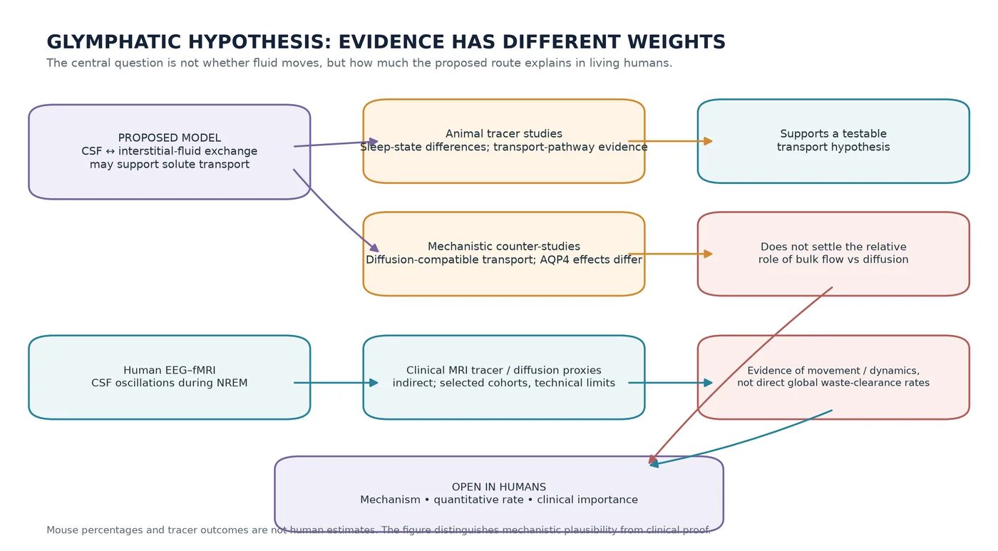
One of the most vivid recent ideas about sleep is that the brain may alter fluid exchange during sleep to aid the removal of waste products. The proposed “glymphatic” model describes cerebrospinal fluid (CSF) entering along periarterial spaces, exchanging with interstitial fluid (ISF) in brain tissue, and helping transport solutes toward perivenous, meningeal, and lymphatic drainage routes. Astrocytic aquaporin-4 (AQP4) channels have been proposed to facilitate this exchange. Some parts of the broader picture—fluid and solute movement, and meningeal lymphatic vessels—are established; the complete, unified periarterial-to-perivenous parenchymal route and its quantitative role remain contested40,46.
The influential early evidence came from mice. A 2012 tracer study reported CSF entry along paravascular spaces and solute clearance along paravenous routes; in its Aqp4-knockout model, measured interstitial tracer clearance was about 70% lower40. A later mouse study reported that natural sleep or anesthesia was associated with roughly a 60% expansion of cortical interstitial-space volume, increased CSF–ISF tracer exchange, and faster clearance of beta-amyloid tracer41. These findings support a sleep-state-dependent transport hypothesis in animals. They do not demonstrate a 60% change in human brain space, a human waste-clearance rate, or that sleep clinically “flushes toxins.”
There is also mechanistic disagreement. A 2017 rodent study found that dextran penetration depended on molecular size in a pattern consistent with diffusion and reported no impairment of measured transport after AQP4 deletion, challenging the claim that convective, AQP4-dependent flow dominates parenchymal solute movement42. This does not prove that convection is absent or that CSF/ISF exchange does not occur. It does mean transport mechanisms, the relative contributions of diffusion and bulk flow, and the interpretation of tracer experiments remain debated46. Injection route, anesthesia, respiration, cardiac pulsation, posture, surgery, and tissue handling can all influence observed tracer distributions46.
Human measurements are suggestive but indirect. A small EEG-fMRI study observed low-frequency, approximately 0.05-hertz CSF inflow oscillations during NREM sleep, coupled to neural and hemodynamic rhythms43. It measured macroscopic oscillations, not clearance of interstitial waste. MRI after intrathecal gadobutrol in clinical participants has shown tracer signal in CSF, selected brain regions, and later cervical lymph nodes44,45. Such observations are consistent with tracer movement through brain-associated pathways, but an administered contrast agent is not endogenous waste, the clinical cohorts are selected, and the method does not establish normal flow direction or quantify physiological clearance in healthy people.
Other human approaches, such as diffusion tensor imaging along perivascular spaces (DTI-ALPS), are proxies rather than direct readings of bulk flow. Their values are affected by imaging plane, head position, and region-of-interest placement, among other technical and biological factors47. Associations between a proxy and age or disease cannot by themselves establish impaired clearance as a cause. The claim that glymphatic dysfunction drives Alzheimer disease or that sleep removes amyloid or tau in humans through this pathway remains unproven47.
The cautious conclusion is not that the hypothesis has been disproved. It is that a productive model of fluid movement and sleep-dependent transport in animals has motivated human investigations, while the human evidence has not yet established brain-wide convective clearance, its rate, or its clinical importance. Sleep clearly involves coordinated changes in brain physiology. Which of those changes materially alter human waste clearance, and by what mechanism, remain questions for careful measurement.
Letting go, without losing the plot
Sleep crosses a threshold when homeostatic need, circadian timing, and distributed neural circuits converge. The threshold is followed not by a blank, but by an organized alternation of stages, changing electrical rhythms, shifting sensory responsiveness, and biological processes whose functions are only partly known. A clock aligns the opportunity for sleep with the environmental day; memory and emotion are modified in ways that depend on what was learned and which outcomes are measured; development and aging alter the architecture without writing a single script for every life.
The science is strongest when it resists tidy slogans. Dreams occur beyond REM, but their adaptive purpose is unresolved. Adenosine participates in sleep-wake regulation, but does not explain the whole homeostatic drive. Sleep loss can impair measured cognition, while many chronic health associations remain vulnerable to confounding and reverse causation. Rodent glymphatic experiments are intriguing, but they are not human proof of overnight brain cleansing. Such qualifications do not diminish sleep’s importance. They show why its architecture is worth studying: it is at once measurable and mysterious, stable enough to recognize, dynamic enough to surprise.
References
- American Academy of Sleep Medicine. “The AASM Manual for the Scoring of Sleep and Associated Events.” 2025 (Version 3 released 2023). https://aasm.org/clinical-resources/scoring-manual/
- American Academy of Sleep Medicine. “The AASM Manual for the Scoring of Sleep and Associated Events: Rules, Terminology and Technical Specifications, Version 2.0.” 2012. https://www.neumosur.net/files/grupos-trabajo/suenio/AASM-Manual-2012.pdf
- Patel, A. K., Reddy, V., and Shumway, K. R. “Physiology, Sleep Stages.” StatPearls, 2024. https://www.ncbi.nlm.nih.gov/books/NBK526132/
- Shirota, A., et al. “Discrepancies in the Time Course of Sleep Stage Dynamics, Electroencephalographic Activity and Heart Rate Variability Over Sleep Cycles in the Adaptation Night in Healthy Young Adults.” 2021. https://www.frontiersin.org/journals/physiology/articles/10.3389/fphys.2021.623401/full
- Borbély, A. A., et al. “The two-process model of sleep regulation: Beginnings and outlook.” 2022. https://pmc.ncbi.nlm.nih.gov/articles/PMC9540767/
- Reichert, C. F., Deboer, T., and Landolt, H.-P. “Adenosine, caffeine, and sleep–wake regulation.” 2022. https://pmc.ncbi.nlm.nih.gov/articles/PMC9541543/
- Saper, C. B., Fuller, P. M., Pedersen, N. P., Lu, J., and Scammell, T. E. “Sleep State Switching.” 2010. https://pmc.ncbi.nlm.nih.gov/articles/PMC3026325/
- Mahoney, C. E., Cogswell, A., Koralnik, I. J., and Scammell, T. E. “The neurobiological basis of narcolepsy.” 2019. https://pmc.ncbi.nlm.nih.gov/articles/PMC6492289/
- Blume, C., Garbazza, C., and Spitschan, M. “Effects of light on human circadian rhythms, sleep and mood.” 2019. https://pmc.ncbi.nlm.nih.gov/articles/PMC6751071/
- Sack, R. L., et al. “Circadian Rhythm Sleep Disorders: Part I, Basic Principles, Shift Work and Jet Lag Disorders.” 2007. https://pmc.ncbi.nlm.nih.gov/articles/PMC2082105/
- St Hilaire, M. A., et al. “The spectral sensitivity of human circadian phase resetting and melatonin suppression to light changes dynamically with light duration.” 2022. https://pmc.ncbi.nlm.nih.gov/articles/PMC9907124/
- Takahashi, M., et al. “Chronotype and social jetlag influence human circadian clock gene expression.” 2018. https://www.nature.com/articles/s41598-018-28616-2
- National Institute for Occupational Safety and Health. “NIOSH Training for Nurses on Shift Work and Long Work Hours: Circadian rhythms and effects of light.” 2020. https://www.cdc.gov/niosh/work-hour-training-for-nurses/longhours/mod2/19.html
- Siclari, F., et al. “Dreaming in NREM Sleep: A High-Density EEG Study of Slow Waves and Spindles.” 2018. https://www.jneurosci.org/content/38/43/9175
- Scarpelli, S., et al. “The Functional Role of Dreaming in Emotional Processes.” 2019. https://pmc.ncbi.nlm.nih.gov/articles/PMC6428732/
- Paller, K. A., Creery, J. D., and Schechtman, E. “Memory and Sleep: How Sleep Cognition Can Change the Waking Mind for the Better.” 2020. https://pmc.ncbi.nlm.nih.gov/articles/PMC7983127/
- Schäfer, S. K., et al. “Sleep’s impact on emotional recognition memory: A meta-analysis of whole-night, nap, and REM sleep effects.” 2020. https://pubmed.ncbi.nlm.nih.gov/32179376/
- van der Helm, E., and Walker, M. P. “Sleep and Emotional Memory Processing.” 2011. https://pmc.ncbi.nlm.nih.gov/articles/PMC4182440/
- Simon, K. C., et al. “Sleep Across the Lifespan: A Neurobehavioral Perspective.” 2025. https://pmc.ncbi.nlm.nih.gov/articles/PMC12174261/
- Paruthi, S., et al. “Recommended Amount of Sleep for Pediatric Populations: A Consensus Statement of the American Academy of Sleep Medicine.” 2016. https://aasm.org/resources/pdf/pediatricsleepdurationconsensus.pdf
- Watson, N. F., et al. “Recommended Amount of Sleep for a Healthy Adult: A Joint Consensus Statement of the American Academy of Sleep Medicine and Sleep Research Society.” 2015. https://pmc.ncbi.nlm.nih.gov/articles/PMC4434546/
- Centers for Disease Control and Prevention. “About Sleep.” 2024. https://www.cdc.gov/sleep/about/index.html
- Mander, B. A., Winer, J. R., and Walker, M. P. “Sleep in Normal Aging.” 2017. https://pmc.ncbi.nlm.nih.gov/articles/PMC5841578/
- Kurina, L. M., et al. “Sleep duration and all-cause mortality: a critical review of measurement and associations.” 2013. https://pmc.ncbi.nlm.nih.gov/articles/PMC3660511/
- Zimmerman, M. E., et al. “The effects of insufficient sleep and adequate sleep on cognitive function in healthy adults.” 2024. https://www.sciencedirect.com/science/article/abs/pii/S2352721823002930
- Alhola, P., and Polo-Kantola, P. “Sleep deprivation: Impact on cognitive performance.” 2007. https://pmc.ncbi.nlm.nih.gov/articles/PMC2656292/
- Schutte-Rodin, S., et al. “Clinical Guideline for the Evaluation and Management of Chronic Insomnia in Adults.” 2008. https://pmc.ncbi.nlm.nih.gov/articles/PMC2576317/
- Kapur, V. K., et al. “Clinical Practice Guideline for Diagnostic Testing for Adult Obstructive Sleep Apnea: An American Academy of Sleep Medicine Clinical Practice Guideline.” 2017. https://pmc.ncbi.nlm.nih.gov/articles/PMC5337595/
- Smith, M. T., et al. “Use of Actigraphy for the Evaluation of Sleep Disorders and Circadian Rhythm Sleep-Wake Disorders: An American Academy of Sleep Medicine Clinical Practice Guideline.” 2018. https://pmc.ncbi.nlm.nih.gov/articles/PMC6040807/
- Allen, R. P., et al. “Restless legs syndrome/Willis-Ekbom disease diagnostic criteria: updated IRLSSG consensus criteria—history, rationale, description, and significance.” 2014. https://pubmed.ncbi.nlm.nih.gov/25023924/
- Baumann, C. R. “A journey through narcolepsy diagnosis: From ICSD 1 to ICSD 3.” 2014. https://pmc.ncbi.nlm.nih.gov/articles/PMC4521660/
- Sobreira-Neto, M. A., et al. “REM sleep behavior disorder: update on diagnosis and management.” 2023. https://pmc.ncbi.nlm.nih.gov/articles/PMC10756822/
- Gauld, C., et al. “A Systematic Review of Sleep–Wake Disorder Diagnostic Criteria Reliability Studies.” 2022. https://pmc.ncbi.nlm.nih.gov/articles/PMC9313077/
- American Academy of Sleep Medicine. “Consumer Sleep Technology: An American Academy of Sleep Medicine Position Statement.” 2018. https://pmc.ncbi.nlm.nih.gov/articles/PMC5940440/
- Edinger, J. D., et al. “Behavioral and Psychological Treatments for Chronic Insomnia Disorder in Adults: An American Academy of Sleep Medicine Clinical Practice Guideline.” 2021. https://pmc.ncbi.nlm.nih.gov/articles/PMC7853203/
- Qaseem, A., et al. “Management of Chronic Insomnia Disorder in Adults: A Clinical Practice Guideline From the American College of Physicians.” 2016. https://www.acpjournals.org/doi/10.7326/M15-2175
- Sateia, M. J., et al. “Clinical Practice Guideline for the Pharmacologic Treatment of Chronic Insomnia in Adults.” 2017. https://pmc.ncbi.nlm.nih.gov/articles/PMC5263087/
- Patil, S. P., et al. “Treatment of Adult Obstructive Sleep Apnea with Positive Airway Pressure: An American Academy of Sleep Medicine Clinical Practice Guideline.” 2019. https://pmc.ncbi.nlm.nih.gov/articles/PMC6374094/
- Auger, R. R., et al. “Clinical Practice Guideline for the Treatment of Intrinsic Circadian Rhythm Sleep-Wake Disorders.” 2015. https://pmc.ncbi.nlm.nih.gov/articles/PMC4582061/
- Iliff, J. J., et al. “A paravascular pathway facilitates CSF flow through the brain parenchyma and the clearance of interstitial solutes, including amyloid β.” 2012. https://pubmed.ncbi.nlm.nih.gov/22896675/
- Xie, L., et al. “Sleep Drives Metabolite Clearance from the Adult Brain.” 2013. https://www.science.org/doi/10.1126/science.1241224
- Smith, A. J., et al. “Test of the ‘glymphatic’ hypothesis demonstrates diffusive and aquaporin- 4-independent solute transport in rodent brain parenchyma.” 2017. https://elifesciences.org/articles/27679
- Fultz, N. E., et al. “Coupled electrophysiological, hemodynamic, and cerebrospinal fluid oscillations in human sleep.” 2019. https://pmc.ncbi.nlm.nih.gov/articles/PMC7309589/
- Ringstad, G., et al. “Glymphatic MRI in idiopathic normal pressure hydrocephalus.” 2017. https://pmc.ncbi.nlm.nih.gov/articles/PMC5841149/
- Eide, P. K., and Ringstad, G. “Magnetic resonance imaging provides evidence of glymphatic drainage from human brain to cervical lymph nodes.” 2018. https://www.nature.com/articles/s41598-018-25666-4
- Hladky, S. B., and Barrand, M. A. “The Brain’s Glymphatic System: Current Controversies.” 2020. https://www.sciencedirect.com/science/article/pii/S0166223620300771
- “The Glymphatic System in Humans: Investigations With Magnetic Resonance Imaging.” 2023. https://pmc.ncbi.nlm.nih.gov/articles/PMC11805491/